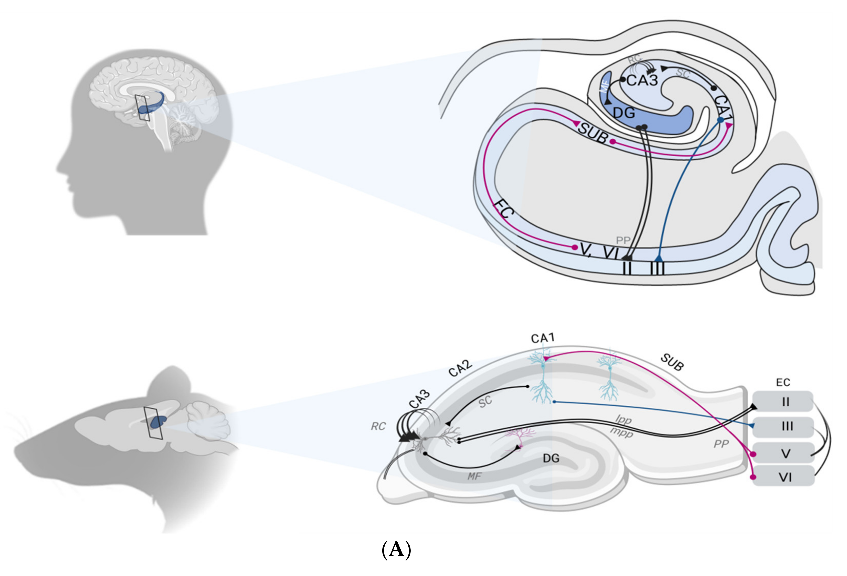
A pulled glass pipette prefilled with virus solution was lowered into the brain, and 165-500 nl of the virus solution was delivered to the targeted brain area using a pressure injection system (NanoJect II, Drummond Scientific Company, Catalog# 3-000-204).
#MOUSE HIPPOCAMPUS ANATOMY SKIN#
The skin along the midline of the skull was opened using a scalpel, and a surgical drill was used to create a small hole in the skull. Anesthesia was maintained for the duration of the surgery by administering isoflurane at 1-2% through a nose cone. Mice were anesthetized with 5% isoflurane and then placed into a stereotaxic alignment instrument (Kopf, model 1900). For ALM experiments, we also injected AAV2-retro-CAG-GFP or AAV2-retro-CAG-tdTomato (Tervo et al., 2016) into wild-type mice. We injected AAV2-retro-EF1a-Cre (Tervo et al., 2016), RV∆GL-Cre (Chatterjee et al., 2018), or CAV-Cre (gift of Miguel Chillon Rodrigues, Universitat Autònoma de Barcelona) (Hnasko et al., 2006) into brains of heterozygous or homozygous Ai14 mice using established procedures (Tasic et al., 2016, 2018). In addition, retrogradely labeled cells were isolated from reporter mice infected with a Cre-dependent virus, or from wild-type mice infected with a reporter virus. Detailed descriptions of recombinase and reporter lines can be found in Transgenic Characterization. Tissue samples were obtained from adult (postnatal day P53-P59) mice, both male and female, carrying one or two recombinase transgenes (Cre, FlpO) and a recombinase-dependent reporter transgene.

#MOUSE HIPPOCAMPUS ANATOMY DRIVER#
For primary visual cortex (VISp) and anterolateral motor cortex (ALM), we sampled additional cells using driver lines that label more specific and rare types. For most brain regions, we isolated labeled cells from pan-GABAergic, pan-glutamatergic, and pan-neuronal transgenic lines. Samples were collected from fine dissections of brain regions from male and female mice. Towards that goal, we have generated a dataset that includes single cells from multiple cortical areas and the hippocampus. Our goal is to quantify the diversity of cell types in the adult mouse brain using large-scale single-cell transcriptomics.


 0 kommentar(er)
